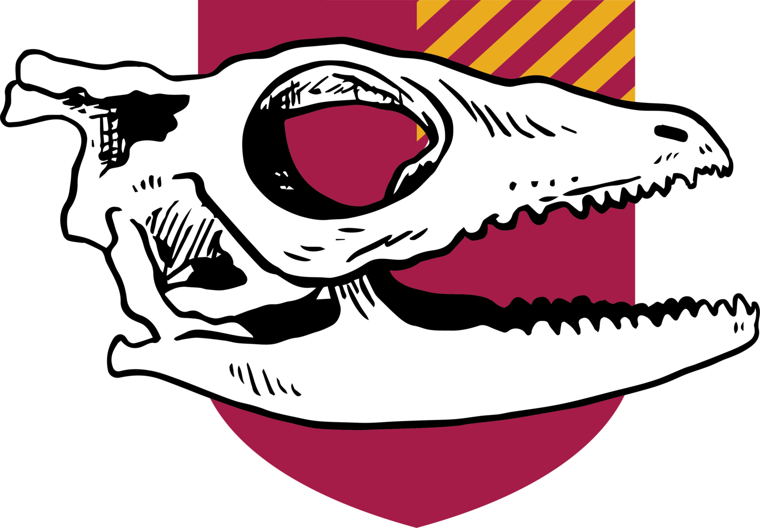
Anolis sagrei skull stages 12-19
Stage 12
The palatine, pterygoid and prootic are first visible ossification centers to form. The palatine is located anteriorly to the pterygoid. The pterygoid forms the posterior-most part of the palate. The prootic, an irregularly shaped bone, forms the lateral sides of the braincase.
Stage 13
Ossification of many bones associated with the circumorbital, temporal, and braincase begins. First visible as a triangularly shaped bone right above the dentary, the premaxilla, located at the anterior front of the snout, and posteriorly to the nasal bone. The maxilla is a large surface bone that makes up the lateral-anterior walls of the orbit of the eye. Framed anteriorly by the maxilla and posteriorly by the postorbital, the jugal begins to ossify, as a large, curved bone that contributes to the lateral part of the orbit. The parietal, seen as two slender angled slivers of bones at this stage, has begun to cover the dorsal posterior portion of the skull. Frontal ossification is seen as a narrow midline that is located dorsally above the palate bones and is coming in contact with posteriorly with the parietal. An irregularly oval shaped bone at this stage, the supratemporal, is located posteriorly at the end of the braincase. It is located to the parietal dorsal laterally. The dentary, a large boomerang shaped bone that starts ossifying at the anterior portion near the snout, and laterally underneath the maxilla and jugal bones, is also coming into contact with the surangular posteriorly and the coronoid dorsal-laterally. The surangular and angular have begun to connect and are located posterior-laterally.
Stage 14
Ossification of the frontal bones begins, and eventually will fuse together to make up the anterior dorsal roofing of the skull. Ossification of circumorbital, skull roof, temporal, braincase, and mandible continues. The nasal bones are irregularly shaped bones that are located anteriorly to the premaxilla, and posteriorly to the prefrontal and frontal circumorbital regions. The maxilla has begun to connect to the jugal. The prefrontal connects posteriorly to the frontal. Another orbital bone, the postorbital bone is a triangular bone that is situated dorsally right above the jugal and anterior-laterally from the squamosum. It also ventrally located under the frontal and parietal. Following the postorbital, the squamosal bone is a curved bone that has begun to connect to the jugal and postorbital anterior- laterally and is connected to the paraoccipital posteriorly. Inside the skull, the palatine has started to connect with the pterygoid and the epipterygoid has appeared as a long and slender bone that is situated ventrally to the pterygoid. The quadrate, a laterally winged shaped bone is positioned ventral-dorsally to the articular, and laterally to the paroccipital. The dentary has begun to fuse with the coronoid and angular. Making up part of the posterior part of the braincase, the paraoccipital has begun to ossify and is located anteriorly to the prootic and the parietal dorsally.
Stage 15
Ossification of the vomer begins here. It is a thin triangular bone, located anterior-ventrally under the premaxilla, laterally to the maxilla, and posteriorly to the palatine. Continued fusion and ossification of jugal, maxilla and prefrontal bones observed. Frontal and parietal bones have connected. Moving to the orbital bones, the lacrimal makes its appearance as part of the orbital surface, connecting anteriorly to the maxilla and prefrontal and posterior-laterally to the jugal. The squamosal and supratemporal have connected. The sphenoid and basioccipital ventrally attached to one another, marks the beginning of ossification of the braincase floor. Continued ossification and fusion of bones observed.
Stage 16
Stage 16-18: The orbitosphenoid, a boomerang shaped bone, hovers vertically in the middle of the skull, underneath the frontal and parietal, anteriorly to the paraoccipital and prootic. It does not come in contact with any bones. Palatine and vomer have connected. In addition, the ectopterygoid begins to ossify, an irregularly shaped bone which connects laterally to the pterygoid and medially to the dentary and coronoid. Paraoccipital and prootic have connected.
Stage 19
Appearance of stapes observed, short horn shaped bones that lie on the posterior-laterally sides of the braincase. Eventually disappears during maturity. All bones have connected, and ossification complete in most bones. The parietal bone eventually ossifies to cover hole after hatching and before maturity.
